Electron microscope images
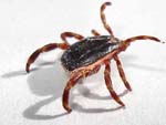
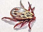
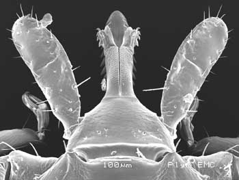
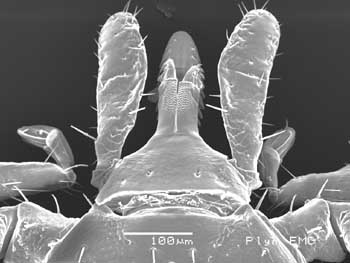
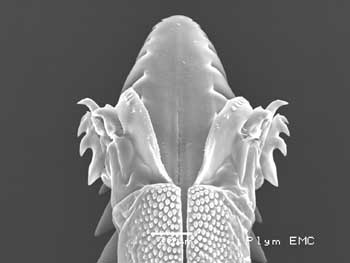
Sheep tick - Ixodes ricinus
NYMPH
Click here for:
larva, nymph,
male, female,
mating ticks and
comparing mouthparts photographs

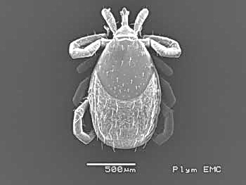
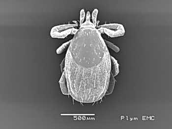
These photos were taken on the JEOL 5600LVSEM
scanning electron microscope at the University of Plymouth Electron Microscopy
Centre on 10th August 2009.
 |
 |
|
Dorsal view, nymph-1 |
Dorsal view nymph-2 |
 |
 |
|
Dorsal view of capitulum, nymph-1
|
Dorsal view of capitulum, nymph-2 |
 |
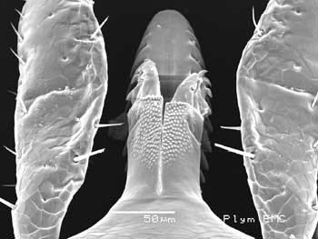 |
Dorsal view of the chelicerae and the tip
of the
hypostome, nymph-1 |
As at left, nymph-2 |
 |
 |
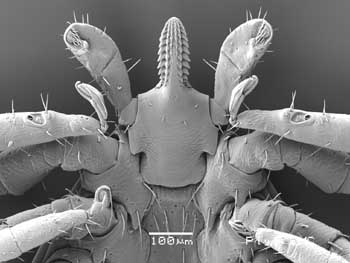
Ventral view of anterior end of nymph,
showing feet,
Haller's organs, pedipalps and hypostome,
also coxae 1 with
spurs |
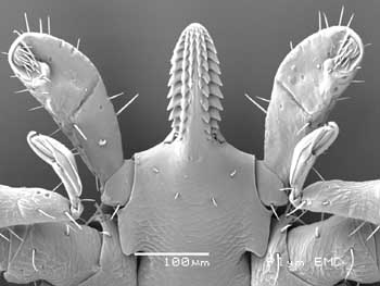
Ventral view of the capitulum
|
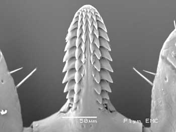 |
Ventral view of the hypostome
showing the recurved barbed denticles |
Acknowledgement: My thanks to the
staff of the University of Plymouth Electron Microscopy Centre, Dr Roy Moate,
Peter Bond and Glenn Harper, for enabling me to take these photographs.
All tick visitors counted by
StatCounter