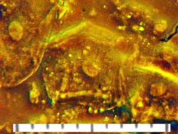
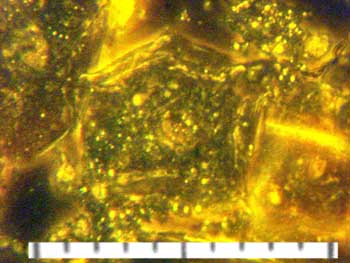
Trial microscopy page
These photos are not tick-based - they are trials
using the borrowed Zeiss Interphako microscope, made in preparation for the
search for Lyme disease-causing bacteria.
The specimen is a scraping of
pavement epithelial cells from the inner lining of the cheek. This is the
material sampled when giving a DNA swab.
The images are taken using
different imaging techniques with the microscope and show a central cell with
another cell at the top left in each image.
The scale bar is 100 micrometers
(mm) with
each division being 10 mm
or 1/100 mm.
It is possible to see 1 mm
diameter particles on the screen, where a 10
mm division
measures 11.4 mm. This means that the 1 mm particles are 1/1.14
mm or 0.877
mm
(<0.001 mm), or 877 nanometers across.
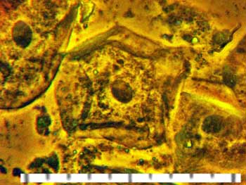
Positive phase contrast - note the dark nucleus in the centre of the cell
(the oval structure).
|
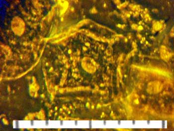
Negative phase contrast - note the light-colour of the nucleus in the
centre of the cell.
|
Coloured phase contrast - note the presence of colours and entities that
are absent from the other micrographs.
|
Dark-field image - note the appearance of granules and vesicles etc. that
are different from the other images.
|
All tick visitors counted by
StatCounter