Measurement
& magnification in microscopes
"Magnification" can often be
meaningless because of the electronic manipulation, resizing etc. of
images on computers and also different computer screen sizes. Magnification
needs to be specifically measured. Often, it is more meaningful to give an
absolute measurement of something seen through a microscope.
Measurements in microscopes are accomplished by calibrating the system using two
items:
-
a stage micrometer - essentially
a small ruler viewed through the microscope, it is an etched scale with divisions. This will
appear larger as magnification is increased.
-
an eyepiece graticule - an etched scale or framework that is located in the
ocular i.e. eyepiece of the microscope. This does not appear larger as magnification is
increased and is therefore a fixed reference.
Wild M5 low-power stereo microscope:
|
|
|
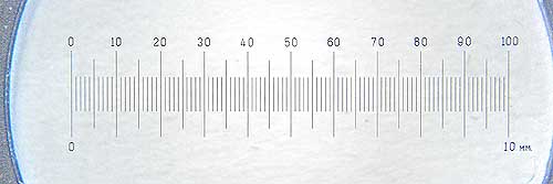
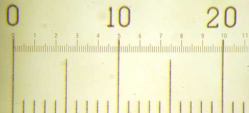
Stage micrometer - scale
for measurement on a "low power" dissecting/stereo microscope
This is a 10 mm scale divided into 100 divisions, where
each small division = 0.1 mm = 100
micrometers = 100 mm. |
|
|
|
Eyepiece measuring graticule in the
Wild M5 stereo microscope
The divisions are arbitrary and need calibrating against a known scale. |
Calibration simply means
recording what the eyepiece graticule/scale divisions cover on the stage micrometer at each
magnification. For practical purposes, it is more accurate to take many
eyepiece divisions, measure them against a length on the stage micrometer
and then calculate what one eyepiece division measures.
|
|
|
Stereo microscope stage
micrometer (large figures) and eyepeiece micrometer (small figures)
photographed at x50. This combines the scales seen in
the two photos above.
100 eyepiece divisions on the small scale cover 2 mm on the large scale,
where "20" = 2,000 micrometers.
Therefore, 1 eyepiece division = 20 micrometers at x50. |
Leitz Orthoplan "high-power"/compound
research microscope
- with Nomarski (DIC - Differential Interference Contrast) and dark-field optics:
|
|
|
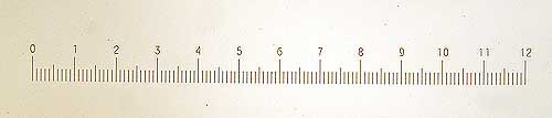
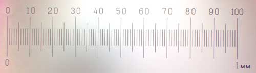
Stage micrometer - scale for measurement on a
high-power microscope
This is a 1 mm scale divided into 100 divisions.
Each small division = 1/100th mm = 0.01 mm = 10
micrometers or 10 mm |
|
|
|
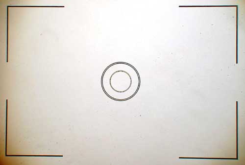
Eyepiece graticule in the
Leitz Orthoplan microscope
(this was originally part of a 35 mm automatic film camera, now obsolete)
The corner lines and ring spacings can be used for measurement. |
Again - calibration simply means
recording what an eyepiece feature covers on the stage micrometer at each
magnification. For accuracy, it is more accurate to take several eyepiece
divisions, measure them against a long length on the stage micrometer and then
calculate what one eyepiece division/feature measures.
|
|
|
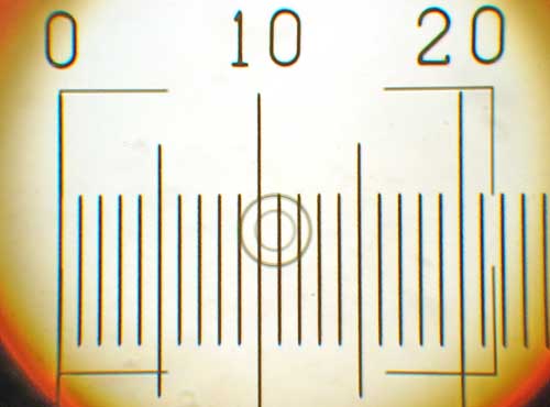
Both Leitz high-power
microscope graticules photographed together at
x400.
The whole photo-field covers 215 micrometers left-to-right, 0.215 mm or
approx. 1/5th mm.
The outer rings cover 35 micrometers
The inner rings cover 20 micrometers
The gap between the 2 rings covers 9 micrometers at x 400.
The magnification of this image?
"0 to 20" = 1/5th mm = 0.2 mm = 200 micrometers covering 142 mm
on a 800x
600 display.
Therefore: 142,000 / 200 = x710
NB - the microscope was set at x400 |
|
|
|
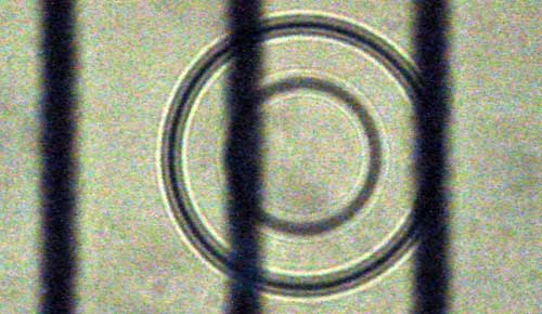
Both high-power microscope
graticules photographed together at
x1000.
The repeat period of the vertical black bars on the stage micrometer is 10
micrometers.
This measures approx. 65 mm on an 800x600 display.
The double outer rings are approx 6.5 mm wide, covering 1 micrometer.
The central dark zone of the outer rings measures approx. 2 mm,
covering 10 x (2/65) = 0.3 micrometers
(this is close to the resolution limit of the best light microscopes. being
about 0.2 micometers).
The magnification on this image?
- on a 800x 600 display
The repeat period of the vertical black bars, representing 10 micrometers,
is 65 mm.
65 mm = 65,000 micrometers, therefore divide by 10 to get 1 micrometer
65,000 / 10 = x6,500 magnification.
|
Another way of measuring things
is to photograph a specimen and then photograph a scale under the same
conditions and compare/superimpose the two without digitally changing sizes.
A further method is to photograph
a specimen and scale at identical magnifications - these can then be
manipulated for measurement in a photo program using separate image layers.
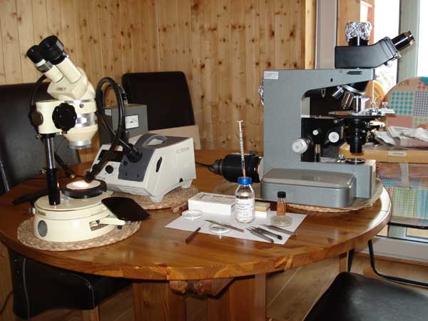
Wild M5 low-power microscope (left)
Leitz Orthoplan microscope (right)
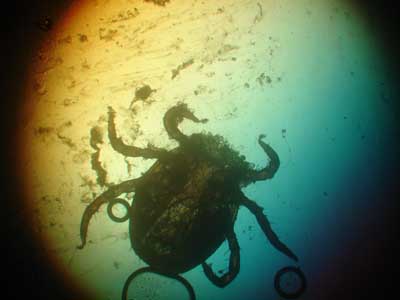
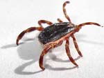
First nymph examined for gut contents, from
Ringmoor Down.
The 'head' and first pair of legs are removed.
The extruded contents are to the left.
All tick visitors counted by
StatCounter